
Anatomy of the Vestibular System: Understanding Your Inner Ear’s Role in Balance
Updated: 10/27/24
Essential Points:
The Vestibular System is Key to Balance: The vestibular system, located in the inner ear, plays a critical role in helping the brain maintain balance by detecting both rotational and linear movements.
Anatomy Breakdown: The system includes structures like the semicircular canals, otolith organs, and hair cells, all working together to sense motion and send information to the brain.
Communication with the Brain: Movement signals from the vestibular system are transmitted to the brain through the vestibulocochlear nerve, where they are integrated with input from the visual and sensory systems to maintain equilibrium.
If you’ve read my article on Three Bodily Balance Systems, you now know that your body’s balance system is controlled by three key components: the visual system, the sensory system, and the vestibular system. As a physical therapist, my focus is primarily on the vestibular system, because it plays such a vital role in helping to compensate for or correct any balance deficits you might experience.
In this article, I’ll take you on a journey through vestibular system anatomy, the part of your body that helps maintain your sense of balance, spatial awareness, and motion detection. Keep in mind that all the structures we discuss exist in both ears, mirroring each other perfectly. Let’s break down each part, from the outside to the inner workings of the ear.
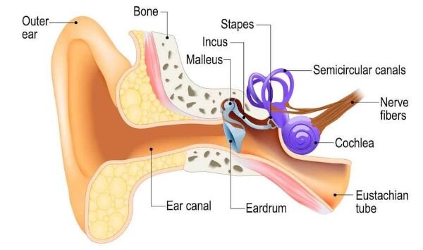
Outer Ear Anatomy: The First Step in Balance and Sound

https://miamiposture.com/wp-content/uploads/2018/09/outer-ear-anatomy.jpg
When you think of your "ear," you’re likely picturing the auricle or pinna, the fleshy part that sticks out from the side of your head. But did you know its design is more than cosmetic? The pinna is perfectly shaped to capture sound waves and funnel them into your ear canal. This canal, about an inch long, channels sound deeper into your ear, where it meets the tympanic membrane, also known as your eardrum.
This section of the ear is what we call the outer ear, and its primary job is to collect sound. However, it also plays a minor but crucial role in your sense of balance by directing vibrations inward.
Middle Ear Anatomy: Where Sound Becomes Movement

https://www.aboutkidshealth.ca/Article?contentid=833&language=English
Just beyond the eardrum, you’ll find the middle ear, home to the smallest bones in your body: the ossicles. These three tiny bones, the malleus (hammer), incus (anvil), and stapes (stirrup), are what convert the sound waves hitting your eardrum into vibrations that can be interpreted by your brain. The stapes, the final bone in the chain, attaches to the oval window, a small opening that leads to the inner ear.
Here’s where things start to get interesting. While this area primarily handles sound transmission, it’s also vital for maintaining pressure equilibrium, something critical for your sense of balance.
Inner Ear: Where Balance Begins
https://www.hpcwire.com/off-the-wire/supercomputing-the-secrets-of-the-inner-ear/#foobox-2/0/inner-ear.jpeg
The inner ear is where both hearing and balance begin to take shape. Inside this part of your ear, you’ll find two major systems: the cochlear apparatus (responsible for hearing) and the vestibular apparatus (responsible for balance). While they share space inside what’s known as the bony labyrinth, their functions are distinctly different.
We’ll just focus on the vestibular apparatus for our purposes, which helps you maintain balance and detect changes in your head’s position. The vestibular apparatus consists of three semicircular canals and two swellings known as otolith organs. Together, these structures detect both rotational movement and linear acceleration, giving your brain the information it needs to help you stay balanced, whether you're walking, running, or just standing still.
Membranous Labyrinth: The Fluid-Filled Inner Chamber

https://www.britannica.com/science/membranous-labyrinth
Within the vestibular portion of the bony labyrinth lies the membranous labyrinth, a delicate, fluid-filled structure suspended in place by perilymph fluid and connective tissue. This inner labyrinth contains five main parts: the three semicircular canals and two otolith organs (utricle and saccule) at their base.
Each semicircular canal ends in a widened region called the ampulla, where specialized hair cells are housed. These hair cells are critical for detecting head movements and sending balance signals to the brain.
Two distinct fluids fill the spaces within the inner ear:
Endolymph fills the membranous labyrinth and plays a direct role in activating hair cells during movement.
Perilymph, surrounding the membranous labyrinth, acts as a cushion within the bony labyrinth.
The separation and unique ionic composition of these fluids ensure proper signaling when the head moves, allowing your vestibular system to maintain balance.
Semicircular Canal Anatomy: Your Body’s Internal Gyroscope

https://www.semanticscholar.org/paper/In-vitro-model-of-a-semicircular-canal%3A-design-and-Obrist-Hegemann/043f0746fe65b47e1094e74c547f8b496f2c48d8/figure/0
Think of the semicircular canals as your body’s internal gyroscope, working to keep your head aligned with your body. These canals come in three orientations, each detecting a different type of rotational movement.
The anterior canal helps you sense nodding movements (like when you say "yes").
The posterior canal detects side-to-side head tilts (like when you lean your ear toward your shoulder).
The lateral canal senses left-to-right rotations (like shaking your head "no").
Each of these canals is filled with fluid and has an enlarged area called the ampulla. Inside the ampulla, special cells called hair cells react to the motion of the fluid when you move your head. These cells are your first line of communication to the brain, telling it which way your head is rotating.
Did You Know?
These canals work in harmony to help you feel full 360-degree movements. So, when you’re spinning in a chair or moving in any direction, your semicircular canals are constantly sending feedback to your brain.
Otolith Organ Anatomy: Detectors of Gravity and Acceleration

https://www.britannica.com/science/otolith
Just below the semicircular canals lie two structures called the otolith organs: the utricle and the saccule. These swellings are key for detecting linear acceleration and gravity. The utricle primarily senses forward and backward movements, while the saccule helps you detect vertical motions, like when you’re riding an elevator.

https://www.britannica.com/science/otolith
What makes the otolith organs unique is their otolithic membranes, which contain tiny calcium carbonate crystals called otoconia. These crystals add weight, allowing the organs to sense the pull of gravity. Whenever you move, the weight of the otoconia shifts, pulling on hair cells embedded in the membrane, which then relay movement information to your brain. This is how your body can tell if you're moving in a straight line or adjusting to gravity.
Vestibular Nerve Anatomy: The Wiring to the Brain

https://mobilephysiotherapyclinic.in/vestibulocochlear-nerve/
Once your hair cells have detected motion, whether rotational or linear, this information is sent to your brain via the vestibular nerve. The vestibular nerve joins with the cochlear nerve to form cranial nerve VIII, also called the vestibulocochlear nerve. This nerve travels back to your brainstem, where the information is processed and integrated with signals from your visual and sensory systems to help you maintain balance.
Final Thoughts: Setting the Stage for Vestibular System Physiology
By understanding the intricate anatomy of the vestibular system, you can better appreciate how your body detects movement and stays balanced. From the fluid-filled semicircular canals to the otolith organs’ clever use of tiny crystals, each part of this system works together to give your brain the information it needs to keep you upright and steady.
In the next article, we’ll dive deeper into vestibular system physiology, how these structures communicate and function on a cellular level to maintain your equilibrium, even in the trickiest of situations.
What’s My Fall Risk Score?

Most people don’t notice their balance declining until something goes wrong.
This 10-minute self-assessment will show you:
• How stable your balance really is
• Where you're most at risk (strength, coordination, or falling ability)
• What to focus on first
No equipment. No guesswork. Just clear answers.
The entirety of this article is based on the information in the following resources.
References
Professional CCM. Vestibular system. Cleveland Clinic. Published October 14, 2024. https://my.clevelandclinic.org/health/body/vestibular-system
Herdman SJ, Clendaniel R. Vestibular rehabilitation. Contemporary Perspectives in R; 2014.
Vestibular system. Kenhub. Published November 3, 2023. https://www.kenhub.com/en/library/anatomy/the-vestibular-system
Thanks for reading about vestibular anatomy! I hope you learned a thing or two after diving into this article. Leave a comment below about what you found most interesting or what you would like me to go more in-depth on in future articles.
If you want to learn more about the vestibular system check out Vestibular Rehabilitation by Susan Herdman. It is THE definitive text on vestibular rehab.
Happy falling!